Overview of Department: Anatomy Museum
Department of Anatomy was established in the year 1998, with a goal to contribute to the vision and mission of the Mamata medical college, in the areas of education, research and service and with an objective to train undergraduate and post graduate students in the fields of gross anatomy, osteology, microscopic anatomy, developmental anatomy, surface anatomy, neuroanatomy, radiological anatomy and also to conduct conferences, workshops and continuing medical education (CME) programs for an updated knowledge on recent advances in human anatomy, for the benefit of students and faculty as well.
The Faculty, Infrastructure and Equipment meet all the requirements of undergraduate and postgraduate teaching & learning and are in accordance with M.C.I and K.N.R.U.H.S Regulations.
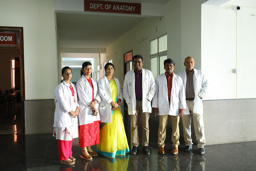
Departmental Faculty:
| S. No. | Faculty Name | AEBAS Attend.ID | Designation | Nature of Employment (Permanent/Contractual) |
| 1 | Dr. Udaya Kumar. P | 11533881 | Professor & HOD | Permanent |
| 2 | Dr. Thondapu Kalpana | 96778912 | Professor | Permanent |
| 3 | Ms. N. Sujatha | 03531716 | Assistant Professor | Permanent |
| 4 | Dr. Indla Edward | 67747031 | Assistant Professor | Permanent |
| 5 | Mr. Morampudi Ravi Kiran | 04970133 | Assistant Professor | Permanent |
| 6 | Ms. Janamala Ratna Priyanka | 32932362 | Assistant Professor | Permanent |
| 7 | Dr. S T R Phanindra | 22231202 | Senior Resident | Permanent |
| 8 | Mr. Tata Venkateswara Babu | 03806199 | Tutor | Permanent |
| 9 | Dr. Katta Bhaavani | 41324231 | Tutor | Permanent |
| 10 | Dr. Kandula Jyoshna | 60314590 | Tutor | Permanent |
| 11 | Dr. Mohammed Zohra Parveen | 59545731 | Tutor | Permanent |
| 12 | Dr. Ravula Bhargavi | 58974872 | Tutor | Permanent |
| 13 | Dr. Swargam Archana | 87146087 | Tutor | Permanent |
| 14 | Dr. Tikkala Chetana | 00978465 | Tutor | Permanent |
| 15 | Dr. Yedlapalli Srija | 41321252 | Tutor | Permanent |
| 16 | Dr. Kokkireni Sathwik | 96495740 | Tutor | Permanent |
| 17 | Dr. Dudam Sahithi | 24311907 | Tutor | Permanent |
UNDER GRADUATE COURSES OFFERED:
MBBS, BDS and B.Sc Nursing
POST- GRADUATE COURSES OFFERED:
MD – Anatomy
TEACHING & LEARNING RESOURCES:
Departmental library is provided with more than 264 books and two desktops with an internet facility.
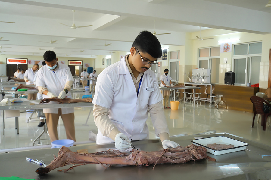
Adequate numbers of cadavers are available for undergraduate and post graduate teaching and for research activities.
OHP & LCD projector, models & charts for gross, microscopic and developmental anatomy teaching.
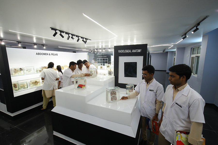
Radiographs: Since Imaging techniques have become increasingly important in diagnosis and treatment, students are introduced to Radiological Anatomy during the course. The X-ray library has meticulously prepared sets of important radiographs, CT scans and MRI films which are demonstrated and discussed with the students.
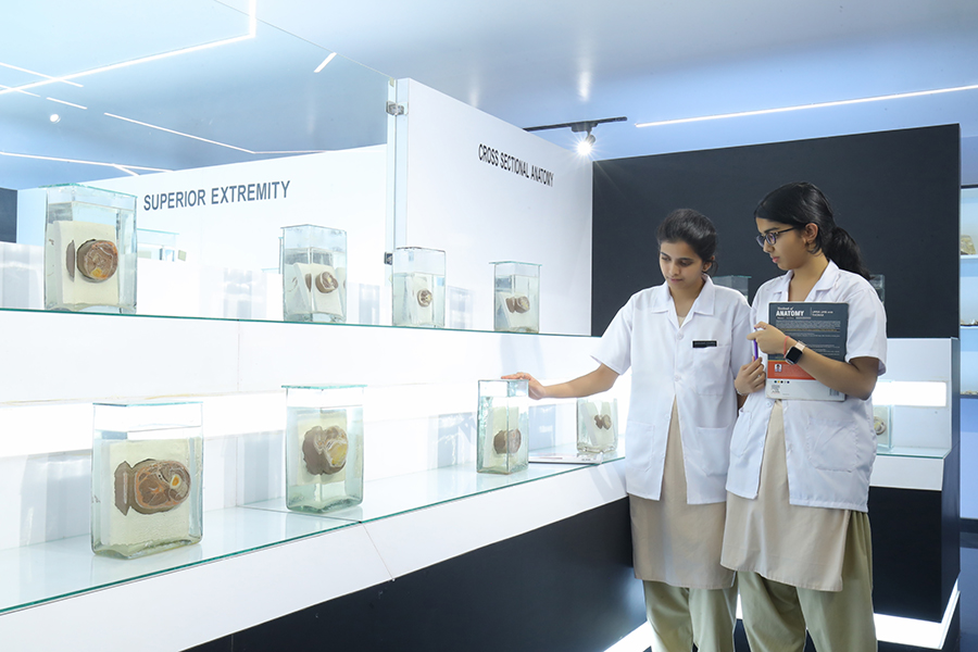
Museum: The Anatomy museum has wide range of dry and wet specimens prepared over the years under the guidance of successive departmental heads. Specimens include gross anatomy, neuroanatomy & embryology, X-ray lobbies with radiographs, genetic charts, skeletons and individual bones. Specimens are displayed system wise in the Museum. Catalogues are placed for quick reference of the specimens.
Bone bank: study of the bones in a region lays down a basic outline of skeletal framework for the further study of the region. Bones taken out of departmental burial ground are cautiously prepared and maintained in the bone bank. The Bone bank is used for teaching osteology purposes and also issued to students for study in the department.
DEPARTMENTAL EQUIPMENT:
Dissection Hall – is provided with
- Two – Body Injectors for embalming procedures,
- Four – Cadaver Storage Tanks to store up to 50 cadavers,
- A Cold Storage (mortuary chamber) facility to preserve 4 dead bodies at a time,
- A Power saw and a Meat Cutting machine for sectioning the bones & soft tissues.
- Dissection hall also has sufficient stretchers, stools, dissection instruments and lockers for students’ utilization and a cordless audio system to communicate to the students.
Histology Lab: is provided with
- Binocular microscope, fitted with a HD camera and LCD projector for demonstration of regular Undergraduate and post graduate histology practical classes.
- Lab is well equipped with 2- Rotary microtomes, one incubator, hot water bath, refrigerator, staining materials and sufficient slide cabinets for preparation and staining of histology slides. H&E and special staining slides are prepared for both UG and PG teaching.
- More than 90 compound microscopes and five dissecting microscopes are available for individual student demonstrations.
FACULTY PAPER PUBLICATIONS:
- Ramanuja Phanindra S.T, A.Raja, K. Yesupadamu, G. Sailaja, D.A.V. S.Sesi. A Cadaveric Study on The Fissure for Ligamentum Teres Hepatis in South-Indian Population. European Journal of Cardiovascular Medicine. Jul-Aug, 2024;14(4):92-99.
- Indla Edward, Ravikiran Morampudi, Prashanth Kumar, Patnaik. Influence Of Patient Anatomical Variability On The Pharmacodynamics Of Intravenous Anaesthetics. An Observational Study. International Journal of Pharmaceutical and Clinical Research. 2024;16(3):1028-1031.2.
- Edward Indla, K V Rajasekar, Bandarupalli Naveen Kumar, S Saravana Kumar, Udaya Kumar P, Suresh Babu Sayana. Modulation of Oxidative Stress and Glycemic Control in Diabetic Wistar Rats: The Therapeutic Potential of Theobroma cacao and Camellia sinensis Diets. Cureus. 2024 Mar 11;16(3):e5598.
- 1. Ravi Kiran Morampudi , Vishali Neelakandan , Bandarupalli Naveen Kumar , Edward Indla. Evaluation of Cognitive and Synaptic Restoration in Diabetic Rats Treated With Emblica officinalis, Clitoria ternatea, Vitamin C, and Metformin. Open Access Original Article . Cureus 16(12):e75866.
- Rajeswara Rao N, Avantika Bamne, Sujatha Nagari, Hemanth Kommuru. Anatomical Variations of the Sciatic Nerve: High Division and Trifurcation and its Clinical Implications International Journal of Medical Science and Current Research. September- October 2024;7(5): 352-357.
- Galenka Vijayasree, Ratnapriyanka Janamala, M.Nagendra Babu. A Study of Morphometic Measurements of Foramen Magnum in Male adult dry skulls. September-2024;14(09):7-10.
- W.R. Reema Rao Incidence & Morphological Variation Rotundum & Foramen Lacerum In 173 Human Skulls From Andhra Pradesh India.
- W.R. Reema Rao Morphological & Morphometic Analysis of Middle Crauial Foramine in adult Human Skulls Implication for Neurological Procedure.
- K.Sangeetha, Shashi Bushan Gollapalli. Anatomical correlation of lung lobes, fissures and pulmonary disorders in cadaveric specimens –An institutional study. Embassy- Research Journal of Medical sciences. 17th oct-2023;17(12)
- Indla E, Rajasekar K, Naveen Kumar B. Neurohistopathological Alterations Induced by Theobroma Cacao and Camellia Sinensis Extracts in Diabetic Male Wistar Rats. Cureus. November 08, 2023;15(11)
- Tirumala Bukkapatnam Ramakrishna, Uma Pokala, Sasikala Kanapalli, Naveen Pokala and Uday Kumar P. Trends in hospitalization and mortality in COVID-19 admitted patients after a single dose of vaccine, Asian Journal of Medical Sciences. Jul 2023;14(7)
- Sujatha Nagari, Hemanth Kommuru, Nithin Kumar Pulluru and Swayam Jothi S. Morphological study & Histological changes that takes Place in human placenta due to nutritional anaemia, Indian Journal of Applied Research, November 2022; 12(11):36-39.
- Vinay G, Benjamin W, Das AK, Raviprasanna KH, Kumar DS, Morphometric study of the distal end of the dry adult humerus of the south Indian population with its clinical applications. Natl J Clin Anat2021;10:70-4.
- Dr. Vinay G, Dr. Nagapraveen Veerapu; A comparison of self-directed learning and lecture methods for teaching embryology among first year medical students; Sch Int J Anat Physiol, Dec 2019; 2(12): 352-355
- Vinay G, Mangala Gowri S R; To assess the relation between finger print pattern and blood groups; Indian Journal of Clinical Anatomy and Physiology 2019;6(4):488–491.
- T. Kalpana, Ramya Sree A , B. Naveen Kumar; Resveratrol reverses brain glutathione system involved neuronal loss after immobilization stress; Int J Anat Res 2019, Vol 7(3.1):6691-6700. ISSN 2321-4287; DOI: https://dx.doi.org/10.16965/ijar.2019.160
- Ramya Sree. A , Udaya Kumar P, Kalpana. T , Vinayaka Naik. I; Morphometric and morphological study of the nutrient foramina in dry human humerus bones of telangana region, Int J Anat Res, 2019;7(1.3):6302-06.